Table of Contents
Anatomical terminology is the shared language that connects physician documentation to accurate code selection. Before a coder can assign a laterality character in ICD-10-CM or select the correct CPT code for an approach, they must understand the standardized system of planes, directional terms, and body positions that every clinician uses to describe where something is located and how a procedure was performed. Misreading a directional term or confusing a body position can lead directly to a wrong code — and to a denied claim.
This foundational vocabulary appears throughout the CPC exam, embedded inside clinical scenarios rather than tested as an isolated topic. A question about a laceration repair will describe the wound using anatomical direction. A question about a joint procedure will reference a specific plane of approach. If you do not fluently understand this terminology, you will misread the scenario before you even reach the coding decision. This guide covers the complete system: anatomical position, the three cardinal planes, the major directional term pairs, and the standard patient positions used in surgery and imaging. For related terminology, see Medical Terminology for Coders.
The Anatomical Position — The Universal Reference Point
Every directional term in medicine is defined relative to a single reference posture called the anatomical position. In the anatomical position, the body stands erect, facing forward, arms at the sides, palms facing forward, and feet flat and facing forward.
This reference point matters because directional terms do not describe where something is relative to gravity or relative to how a patient happens to be lying on an exam table — they describe location relative to this fixed standard posture. A wound on the “anterior” thigh is anterior regardless of whether the patient is lying on their back, their side, or standing up. Understanding that the anatomical position is a fixed reference frame, not a literal description of the patient’s posture during the encounter, prevents a common misreading of clinical documentation.
The Three Anatomical Planes
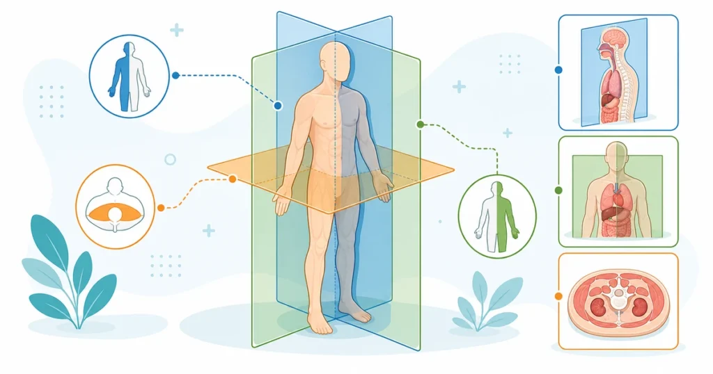
Anatomical planes are imaginary flat surfaces that pass through the body and divide it into sections. They are used constantly in radiology reports, surgical approach descriptions, and imaging orders — all of which a coder must interpret correctly.
| Plane | Also Called | Divides the Body Into | Common Use |
|---|---|---|---|
| Sagittal | Lateral plane | Left and right portions | MRI/CT slices; describes midline structures |
| Coronal | Frontal plane | Front (anterior) and back (posterior) portions | Standard imaging plane; used in describing fracture displacement |
| Transverse | Axial or horizontal plane | Upper (superior) and lower (inferior) portions | Cross-sectional CT/MRI imaging; most common axial imaging plane |
The Sagittal Plane
The sagittal plane runs vertically and divides the body into left and right sections. The midsagittal (or median) plane runs directly through the midline, creating exactly equal left and right halves. Any sagittal plane offset from the midline is called a parasagittal plane, producing unequal left and right portions. Radiology reports frequently reference sagittal imaging when describing spinal alignment or midline structures such as the corpus callosum.
The Coronal Plane
The coronal plane, also called the frontal plane, runs vertically and divides the body into anterior (front) and posterior (back) sections. This plane is commonly referenced in orthopedic imaging, particularly when describing the displacement direction of a fracture fragment relative to the front or back of a limb.
The Transverse Plane
The transverse plane runs horizontally and divides the body into superior (upper) and inferior (lower) sections. It is also called the axial or horizontal plane. Cross-sectional CT and MRI imaging is most often acquired in the transverse plane, producing the familiar “slice” images that radiologists review from the bottom of the body upward.
Directional Terms — Paired Opposites
Directional terms always come in opposing pairs, and each pair describes a relationship between two points relative to the anatomical position. Understanding these as pairs — rather than memorizing them individually — makes them far easier to apply correctly in a clinical scenario.
| Term | Meaning | Opposite Term | Example |
|---|---|---|---|
| Anterior (ventral) | Toward the front of the body | Posterior (dorsal) | The sternum is anterior to the spine |
| Posterior (dorsal) | Toward the back of the body | Anterior (ventral) | The scapula is posterior to the ribs |
| Superior (cephalad) | Toward the head, above | Inferior (caudad) | The heart is superior to the diaphragm |
| Inferior (caudad) | Toward the feet, below | Superior (cephalad) | The stomach is inferior to the esophagus |
| Medial | Toward the midline of the body | Lateral | The nose is medial to the eyes |
| Lateral | Away from the midline | Medial | The ears are lateral to the eyes |
| Proximal | Closer to the point of attachment/trunk | Distal | The elbow is proximal to the wrist |
| Distal | Farther from the point of attachment/trunk | Proximal | The fingers are distal to the wrist |
| Superficial | Closer to the surface of the body | Deep | The skin is superficial to the muscle |
| Deep | Farther from the surface of the body | Superficial | The bone is deep to the muscle |
| Ipsilateral | On the same side of the body | Contralateral | An ipsilateral lymph node biopsy is on the same side as the tumor |
| Contralateral | On the opposite side of the body | Ipsilateral | A contralateral finding is on the side opposite the original condition |
Proximal and Distal Apply Only to the Limbs
A frequent point of confusion is that proximal and distal describe position along the length of a limb or an elongated structure — relative to its point of attachment to the trunk — while superior and inferior describe overall vertical position on the body as a whole. The proximal humerus is the end near the shoulder, and the distal humerus is the end near the elbow. These terms would not typically be used to describe, for example, the position of the liver relative to the head.
Why Laterality Terms Matter for ICD-10-CM
Ipsilateral and contralateral terminology directly affects ICD-10-CM code selection because many code categories include laterality as part of the code structure — right, left, or bilateral. When documentation describes a “contralateral” finding, the coder must determine which side that refers to based on the side already established for the primary condition, then assign the correct laterality character. Getting the ipsilateral/contralateral relationship backward produces a code for the wrong side of the body, which is both a clinical accuracy error and, in many payer systems, a claim-rejecting error.
Regional and Combined Directional Terms
Beyond the primary directional pairs, several regional and combination terms appear regularly in documentation.
Anteroposterior (AP) — from front to back; commonly used to describe the direction of an imaging beam or a measurement taken through the body from front to back.
Posteroanterior (PA) — from back to front; the standard direction for a routine chest X-ray, distinguished from AP views, which are typically used for portable or bedside imaging.
Anterolateral — a combination describing a position that is both toward the front and toward the side, such as the anterolateral thigh, a common donor site for skin flaps and a site referenced in wound repair coding.
Posteromedial — toward the back and toward the midline, frequently used in orthopedic documentation describing fracture fragment displacement.
Palmar (volar) — referring to the palm side of the hand.
Plantar — referring to the sole of the foot.
Dorsal (of the hand or foot) — referring to the back of the hand or the top of the foot; note that “dorsal” has a different practical meaning depending on whether it is applied to the trunk (where it means posterior) versus the hand or foot (where it means the top/back surface).
Standard Patient Positions
Body positions describe how a patient is physically oriented during an examination, procedure, or imaging study. Unlike directional terms, which are fixed relative to anatomical position, body positions describe the patient’s actual posture at the time of the encounter — and this distinction is frequently tested.
| Position | Description | Common Use |
|---|---|---|
| Supine | Lying flat on the back, face up | Most common surgical position; standard for abdominal and chest procedures |
| Prone | Lying flat on the front, face down | Spinal surgery, posterior procedures |
| Lateral decubitus | Lying on one side (right or left) | Thoracic surgery, certain endoscopic procedures |
| Lithotomy | Supine with hips and knees flexed, legs supported in stirrups | Gynecological, urological, and rectal procedures |
| Trendelenburg | Supine with the head lower than the feet (tilted) | Certain abdominal and pelvic surgeries; improves visualization |
| Fowler’s | Semi-sitting, with the head of the bed elevated | Respiratory support, some upper body procedures |
| Sims’ | Lying on the left side with the right knee and thigh drawn up | Rectal exams, enema administration |
Why Position Terminology Matters for Coding
Certain CPT codes explicitly specify or imply the position required for the procedure, and operative reports frequently document the position as part of establishing that the correct technique was used. While the position itself is rarely the deciding factor in code selection, it functions as a cross-check: if an operative report describes a lithotomy position for what should be a routine abdominal laparotomy, the documentation may be internally inconsistent and worth flagging for query. Recognizing standard position terminology also helps a coder read and interpret an operative report efficiently rather than stumbling over unfamiliar vocabulary.
Body Cavities and Regions
Anatomical terminology also divides the body into cavities and regions, which coders encounter when interpreting documentation about the location of an internal finding, effusion, or procedure.
The dorsal cavity contains the cranial cavity (brain) and the spinal cavity (spinal cord). The ventral cavity contains the thoracic cavity (heart, lungs, mediastinum) and the abdominopelvic cavity, which is further divided into the abdominal cavity (stomach, intestines, liver, spleen, kidneys) and the pelvic cavity (bladder, reproductive organs, rectum).
The abdomen is commonly divided into four quadrants (right upper, left upper, right lower, left lower) for general clinical description, and into nine regions (right hypochondriac, epigastric, left hypochondriac, right lumbar, umbilical, left lumbar, right iliac, hypogastric, left iliac) for more precise anatomical localization, particularly in surgical and radiology documentation. Quadrant terminology appears directly in signs and symptoms coding — for example, right lower quadrant abdominal pain has its own specific ICD-10-CM code.
How the CPC Exam Tests This Topic
Pattern 1 — Directional Term Application
The question describes the relationship between two anatomical structures (“the radius is ___ to the ulna”) and asks you to select the correct directional term from a list of answer choices. These questions test whether you can correctly apply proximal/distal, medial/lateral, and anterior/posterior relationships to specific anatomical structures.
Pattern 2 — Laterality and Contralateral/Ipsilateral Reasoning
The scenario describes a primary condition on one side of the body and a subsequent finding described as “contralateral” or “ipsilateral.” You must determine the correct side for the ICD-10-CM laterality character based on the described relationship, not simply default to “right” or “unspecified.”
Pattern 3 — Position Identification from an Operative Report
The scenario quotes language from an operative report describing the patient’s position (for example, “patient placed in lithotomy position”) and asks what type of procedure this position is most consistent with, or asks you to identify the correct position term from a description of the patient’s posture.
Pattern 4 — Plane Identification in Imaging Orders
The question describes an imaging order or a radiology report referencing a specific plane (sagittal, coronal, transverse) and asks what that plane shows or how the body is divided by it.
Common Mistakes
Confusing proximal/distal with superior/inferior. Proximal and distal apply specifically to structures along a limb relative to the trunk. Superior and inferior describe overall vertical position on the body. Using the wrong pair when describing a limb structure is a frequent error.
Misreading dorsal on the hand or foot. Dorsal means posterior when applied to the trunk, but on the hand and foot it refers to the top/back surface — the opposite side from palmar or plantar. Applying the trunk definition to the hand or foot produces the wrong directional meaning.
Reversing ipsilateral and contralateral. These terms are easy to swap under time pressure. Always re-read the sentence carefully to confirm which side is being referenced relative to the originally stated side.
Assuming body position dictates the plane of the procedure. A patient’s physical position (supine, prone, lateral decubitus) is not the same as an anatomical plane (sagittal, coronal, transverse). These are two separate systems of terminology that describe different things and should not be conflated.
Treating anatomical position as the patient’s literal posture during the encounter. Directional terms are always defined relative to the fixed anatomical position, not the position the patient happens to be in during the exam or procedure.
5 quick questions drawn from this guide. Click an answer to check it — explanations appear as you go.
1. The plane that divides the body into left and right portions is the:
2. A transverse (axial) plane divides the body into:
3. ‘Distal’ means:
4. A patient lying face-down is in the ______ position.
5. The coronal (frontal) plane divides the body into:
Frequently Asked Questions
What is the anatomical position?
The anatomical position is the standard reference posture used to define all directional terms in medicine: standing erect, facing forward, arms at the sides, palms facing forward, and feet flat and facing forward. Directional terms like anterior, posterior, medial, and lateral are always defined relative to this fixed position, not the patient’s actual posture during an encounter.
What is the difference between proximal and distal?
Proximal means closer to the point of attachment to the trunk, and distal means farther from that point of attachment. These terms apply specifically to structures along a limb — for example, the proximal humerus is near the shoulder, while the distal humerus is near the elbow. They are not typically used to describe overall vertical position on the body.
What are the three anatomical planes?
The three anatomical planes are the sagittal plane (divides the body into left and right), the coronal or frontal plane (divides the body into front and back), and the transverse plane (divides the body into upper and lower). These planes are used throughout radiology to describe imaging orientation and are frequently referenced in CT and MRI reports.
What does contralateral mean in medical coding?
Contralateral means occurring on the opposite side of the body from a previously referenced condition or structure. This term directly affects ICD-10-CM laterality coding — when documentation describes a contralateral finding, the coder must assign the correct right, left, or bilateral character based on which side the original condition was on, not simply default to the same side.
What is the difference between supine and prone position?
Supine means lying flat on the back, face up, and is the most common surgical position used for abdominal and chest procedures. Prone means lying flat on the front, face down, and is typically used for spinal surgery and other posterior procedures. Both are patient positions during an encounter, distinct from anatomical directional terms.