Advertisement
Table of Contents
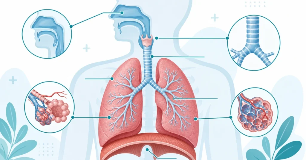
The respiratory system is fundamental to life, delivering oxygen to the body and removing carbon dioxide. As a coder, you’ll encounter respiratory conditions constantly — from simple upper respiratory infections to complex chronic obstructive pulmonary disease (COPD) and acute respiratory failure. Understanding respiratory anatomy and terminology is essential for accurate ICD-10-CM coding.
Upper Airway Structures
Air enters the body through the upper airway, which includes several structures:
- Nasal Cavity: First air pathway, filters and warms air
- Pharynx (Throat): Common pathway for air and food. Divided into nasopharynx (upper), oropharynx (middle), and laryngopharynx (lower)
- Larynx (Voice Box): Contains vocal cords, enables speech, protects airway
- Epiglottis: Flap that covers larynx during swallowing to prevent food from entering lungs
- Trachea (Windpipe): Tube that carries air to the lungs
Lower Airway and Lungs
The Bronchial Tree
Below the trachea, the airway branches like a tree:
- Primary Bronchi: Trachea splits into left and right main bronchi
- Secondary Bronchi: Right main bronchus splits into three lobes; left into two lobes
- Bronchioles: Smallest branches, lead to alveoli
- Alveoli: Tiny air sacs where gas exchange occurs (oxygen in, carbon dioxide out)
⭐ For Coders: The right lung has three lobes (upper, middle, lower) while the left lung has two lobes (upper, lower). When coding pneumonia or other lung conditions, you must specify which lobe is affected. This detail matters for severity and treatment planning.
Pleura and Surrounding Structures
The lungs are surrounded by protective membranes:
- Visceral Pleura: Inner layer adhering to lungs
- Parietal Pleura: Outer layer lining chest wall
- Pleural Space: Area between layers, normally contains small amount of fluid for lubrication
- Mediastinum: Central compartment of chest containing heart, esophagus, and major vessels
Essential Respiratory Terminology
Conditions of the Upper Airway
- Rhinitis: Inflammation of nasal mucosa, causes runny nose
- Sinusitis: Inflammation of sinuses
- Pharyngitis: Sore throat, inflammation of pharynx
- Laryngitis: Inflammation of larynx, causes hoarseness
- Croup: Viral infection of larynx, characteristic barking cough in children
- Epiglottitis: Serious infection of epiglottis, can block airway
Conditions of the Lower Airway and Lungs
- Bronchitis: Inflammation of bronchi, productive cough with mucus
- Pneumonia: Infection of alveoli, fills with fluid/pus, impairs gas exchange
- Asthma: Chronic airway inflammation, bronchospasm, wheezing
- COPD (Chronic Obstructive Pulmonary Disease): Emphysema and chronic bronchitis, progressive airflow obstruction
- Emphysema: Destruction of alveoli, loss of elastic recoil
- Pulmonary Fibrosis: Scarring of lung tissue, progressive stiffness
- Atelectasis: Collapse of alveoli, reduced gas exchange
Pleural and Respiratory Emergencies
- Pneumothorax: Air in pleural space, causes lung collapse
- Hemothorax: Blood in pleural space
- Pleural Effusion: Fluid buildup in pleural space (exudate or transudate)
- Pleurisy (Pleuritis): Inflammation of pleura, severe chest pain with breathing
- Respiratory Failure: Inability to oxygenate or ventilate, types include Type I (hypoxemic) and Type II (hypercapnic)
- Acute Respiratory Distress Syndrome (ARDS): Severe respiratory failure with bilateral infiltrates
Gas Exchange and Respiratory Function
| Term | Definition | Clinical Significance |
|---|---|---|
| Ventilation | Movement of air in and out of lungs | Impaired in COPD, asthma, neuromuscular disease |
| Perfusion | Blood flow to lungs for gas exchange | Impaired in pulmonary embolism, heart failure |
| Gas Exchange | Transfer of O2 and CO2 at alveoli | Impaired in pneumonia, fibrosis, ARDS |
| Oxygenation | Oxygen loading onto hemoglobin | Measured by SpO2 and PaO2 |
| Ventilation/Perfusion (V/Q) | Matching of ventilated air to perfused blood | V/Q mismatch causes hypoxia |
Stages of COPD — A Practical Coding Example
COPD is one of the most common respiratory conditions coded. Understanding COPD stages helps you code severity accurately:
GOLD Stage 1-2
- Mild to moderate airflow limitation
- FEV1 50-80% predicted
- Few symptoms
- Code: Mild to unspecified
GOLD Stage 3-4
- Severe to very severe obstruction
- FEV1 <30% predicted
- Significant dyspnea
- Code: Severe or with complications
Common Respiratory Coding Mistakes
❌ Mistake #1: Coding “upper respiratory infection” without specifying which structure (rhinitis, sinusitis, pharyngitis). The specific site must be identified.
❌ Mistake #2: Forgetting to code lobar pneumonia specificity. If clinical note specifies right lower lobe or left upper lobe pneumonia, this detail must be captured in the code.
❌ Mistake #3: Confusing COPD without exacerbation from COPD with acute exacerbation. Exacerbations get additional codes and change severity.
Why Respiratory Anatomy Matters
Understanding respiratory anatomy helps you:
- Recognize when clinical documentation is complete or missing required specificity
- Understand why certain respiratory conditions are more serious than others
- Code respiratory failure severity appropriately (hypoxic vs hypercapnic)
- Understand procedures like bronchoscopy, intubation, and mechanical ventilation
Advertisement