Table of Contents
- Benign vs. Malignant Lesion Excision Codes
- Measuring Lesion Size for Code Selection
- Integumentary Excision Code Ranges
- Excision vs. Shaving vs. Destruction
- Coding the Closure Separately
- Coding Multiple Lesions
- Re-Excision for Positive or Close Margins
- Mohs Micrographic Surgery
- Anatomic Location Groupings and Why They Matter
- Documentation Requirements for Accurate Coding
- How the CPC Exam Tests Skin Lesion Excision Coding
- Common Mistakes
- Coordinating Excision Coding With Pathology Turnaround
- Frequently Asked Questions
Skin lesion excision is one of the most frequently performed outpatient procedures and one of the most commonly tested integumentary topics on the CPC exam, because correct code selection depends on several interacting variables: the lesion’s behavior (benign or malignant), its measured size, and the type of closure performed afterward. Missing any one of these details in an operative note leads to an incorrect code, even when the coder has otherwise correctly identified the procedure performed. This guide walks through the full excision coding process, building on the closure concepts introduced in Wound Repair Coding and the general surgical coding approach in Surgery Coding for Beginners. Mastering this topic also reinforces the broader documentation-reading discipline that carries over into nearly every other integumentary and surgical coding scenario tested on the exam.
Benign vs. Malignant Lesion Excision Codes
The single most important distinction in lesion excision coding is whether the lesion is benign or malignant, since CPT maintains entirely separate code series for each. Benign lesion excision codes are used when pathology confirms the lesion is non-cancerous, or when the physician’s clinical impression is benign and no malignancy is subsequently identified. Malignant lesion excision codes are used when pathology confirms cancer, such as basal cell carcinoma, squamous cell carcinoma, or melanoma. Because the pathology report often is not available at the time of coding, many practices code initially based on the pre-excision clinical impression and adjust the code if a later pathology report changes the diagnosis from what was initially assumed.
Measuring Lesion Size for Code Selection
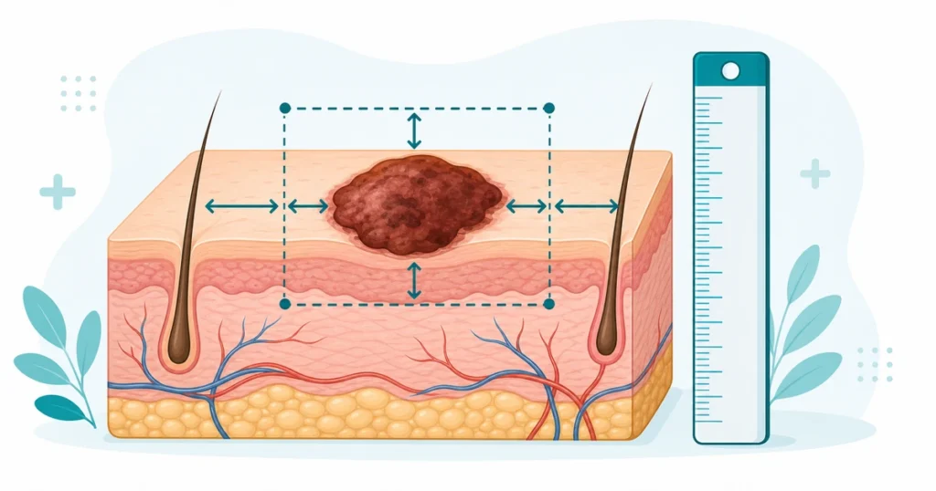
Both benign and malignant excision codes are organized by size, and correctly measuring that size is essential to selecting the right code. The size used for coding purposes is not simply the visible diameter of the lesion itself — it is the lesion’s greatest diameter plus the narrowest margin required for complete excision, added together and measured before the excision is performed. For example, a 1.0 cm lesion excised with a 0.5 cm margin on each side yields a total excised diameter of 2.0 cm for coding purposes, not 1.0 cm. This measurement must be documented by the surgeon at the time of the procedure, since the specimen shrinks after removal and fixation, making a post-excision pathology measurement unreliable for code selection.
Integumentary Excision Code Ranges
| Category | Approximate Range | Organized By |
|---|---|---|
| Excision, benign lesion | 11400–11471 | Anatomic location and size |
| Excision, malignant lesion | 11600–11646 | Anatomic location and size |
| Shaving of epidermal/dermal lesion | 11300–11313 | Anatomic location and size |
| Destruction, benign lesion | 17000–17250 | Anatomic location, method, and number of lesions |
| Destruction, malignant lesion | 17260–17286 | Anatomic location and size |
Both the benign and malignant excision series are further divided by anatomic location groupings, such as trunk/arms/legs versus scalp/neck/hands/feet/genitalia versus face/ears/eyelids/nose/lips/mucous membrane, since these regions carry different code ranges reflecting differences in typical procedure complexity by body area.
Excision vs. Shaving vs. Destruction
New coders frequently confuse excision with two related but distinct procedures: shaving and destruction. An excision is a full-thickness removal of the lesion through the dermis, typically requiring some form of closure. A shave removal takes a superficial, tangential slice of tissue without a full-thickness cut and typically does not require sutured closure. Destruction refers to methods that eliminate the lesion without removing an intact specimen for pathology, such as cryosurgery, laser ablation, or electrodesiccation. Each of these three approaches has its own separate code series, and the operative note’s description of the technique used — not just the fact that “a lesion was removed” — determines which series applies.
Coding the Closure Separately
Because excision codes describe only the removal of the lesion itself, the repair or closure performed afterward is frequently reportable as a separate code, following the same simple, intermediate, and complex repair classifications covered in Wound Repair Coding. A simple, single-layer closure is often considered included in the excision code and not separately reported, while an intermediate closure (requiring layered closure of the subcutaneous tissue) or a complex closure (requiring extensive undermining, debridement, or other advanced techniques) is typically separately reportable in addition to the excision code. Coders should always check the operative note for the specific closure technique used and apply the general rule that more extensive repairs beyond simple closure are usually billed as an additional code.
Coding Multiple Lesions
When multiple lesions are excised during the same operative session, each lesion is coded separately based on its own size, location, and benign/malignant status, rather than being combined into a single code representing the total area treated. If four benign lesions of different sizes on the arm are excised during the same visit, four separate excision codes are reported, each reflecting the specific size of that individual lesion. Modifier -59, or a more specific X-modifier, along with anatomic modifiers when applicable, may be needed to indicate that each code represents a distinct lesion at a separate site rather than a duplicate billing of the same procedure.
Re-Excision for Positive or Close Margins
When pathology reveals that a malignant lesion was not completely removed with adequate margins on the first excision, a second, separate excision procedure to obtain clear margins is coded as its own distinct excision using the appropriate malignant lesion excision code for the new procedure, based on the size of tissue excised during that second procedure. This is coded as a new procedure rather than as a repeat or correction of the original code, since it represents an entirely separate surgical encounter addressing the positive margin finding.
Mohs Micrographic Surgery
Mohs micrographic surgery is a specialized technique for removing certain skin cancers in which the surgeon acts as both surgeon and pathologist, removing a thin layer of tissue and examining it under a microscope in real time before determining whether additional tissue needs to be removed. Mohs surgery has its own distinct code series, separate from the standard malignant lesion excision codes, and is coded based on the anatomic location and the number of tissue stages examined during the procedure, rather than by lesion size in the same way standard excision codes are structured. Recognizing that a documented Mohs procedure should never be coded using the standard malignant excision codes is an important distinction tested on the exam.
Anatomic Location Groupings and Why They Matter
Both benign and malignant excision code series divide the body into distinct anatomic groupings rather than treating all skin surfaces identically, and choosing the wrong grouping produces an incorrect code even when the size and benign/malignant status are correctly identified. Trunk, arms, and legs form one grouping; scalp, neck, hands, feet, and genitalia form a second; and face, ears, eyelids, nose, lips, and mucous membrane form a third, more complex grouping reflecting the greater technical difficulty and cosmetic sensitivity of procedures in those areas. A lesion of identical size and behavior excised from the back versus the eyelid will use entirely different codes because of this anatomic grouping structure, so coders must always confirm the precise anatomic site documented rather than assuming a single universal code applies regardless of location.
Documentation Requirements for Accurate Coding
Because so much of lesion excision coding depends on precise measurements and technique descriptions, thorough operative documentation is essential to correct coding, and incomplete documentation is a frequent source of coding delays and denials. The operative note should clearly state the lesion’s pre-excision diameter, the margin taken, the anatomic location, whether the lesion was clinically benign or suspicious for malignancy, the technique used (excision, shave, or destruction), and the type of closure performed. When any of these elements is missing from the documentation, coders may need to query the physician for clarification rather than guessing, since an incorrect assumption on any single element can produce a code that does not match what was actually performed.
How the CPC Exam Tests Skin Lesion Excision Coding
Pattern 1 — Calculating Total Excised Diameter
A scenario provides both the lesion size and the margin size, testing whether you correctly add the margin to the lesion diameter to determine the size used for code selection, rather than using the lesion size alone.
Pattern 2 — Distinguishing Excision, Shave, and Destruction
A scenario describes a lesion removal technique, testing whether you correctly identify which of the three code series (excision, shave, destruction) applies based on the specific technique documented.
Pattern 3 — Coding the Closure Separately
A scenario describes an excision followed by an intermediate or complex repair, testing whether you recognize that this closure is separately reportable in addition to the excision code, unlike a simple single-layer closure.
Pattern 4 — Coding Multiple Lesions at Different Sites
A scenario describes several lesions excised during the same session, testing whether you correctly report a separate code for each lesion based on its individual size and location, with appropriate modifiers.
Common Mistakes
Using the lesion diameter alone without adding the margin. The size used for code selection is the lesion diameter plus the narrowest margin required for complete excision, not the visible lesion size alone.
Confusing excision, shave, and destruction codes. These represent three distinct code series based on the specific technique documented, not interchangeable terms for lesion removal.
Failing to code a separately reportable closure. Intermediate and complex repairs performed after excision are frequently separately billable and should not be assumed to be bundled into the excision code.
Coding multiple lesions as a single combined procedure. Each excised lesion is coded individually based on its own size and location.
Coding Mohs surgery with standard excision codes. Mohs micrographic surgery has its own dedicated code series based on anatomic location and number of stages, not lesion size.
Coding based on the pathology-measured specimen size rather than the surgeon’s pre-excision measurement. Specimens shrink after removal and fixation, so the surgeon’s documented pre-excision measurement, including margins, is the correct basis for code selection.
Coordinating Excision Coding With Pathology Turnaround
Because the definitive benign-versus-malignant status of a lesion often depends on a pathology report that returns days after the procedure, practices need a clear internal process for updating claims when the pathology diagnosis differs from the initial clinical impression used at the time of coding. If a lesion coded as benign at the time of surgery is later confirmed malignant on pathology, the claim may need to be corrected to the appropriate malignant excision code once the report is available, rather than left as originally submitted. Establishing a routine check of pending pathology results against previously coded excisions helps ensure that final coding accurately reflects the confirmed diagnosis rather than only the pre-procedure clinical impression.
5 quick questions drawn from this guide. Click an answer to check it — explanations appear as you go.
1. CPT excision codes are chosen first by:
2. The measured “excised diameter” equals:
3. A simple, intermediate, or complex repair of the excision wound is:
4. Excision codes are selected using the size in:
5. For multiple lesion excisions, you generally:
Frequently Asked Questions
How do you determine the correct size for skin lesion excision coding?
The size used for coding is the lesion’s greatest diameter plus the narrowest margin required for complete excision, measured by the surgeon before the excision is performed. This combined measurement, not the visible lesion size alone, determines the correct code.
What is the difference between excision, shave removal, and destruction of a skin lesion?
Excision is a full-thickness removal through the dermis, typically requiring closure. Shave removal is a superficial, tangential slice without full-thickness cutting. Destruction eliminates the lesion using methods like cryosurgery or laser ablation without producing an intact specimen. Each has its own separate CPT code series.
Is wound closure after a lesion excision coded separately?
A simple, single-layer closure is generally included in the excision code. An intermediate closure requiring layered repair or a complex closure requiring extensive techniques is typically separately reportable in addition to the excision code.
How do you code excision of multiple skin lesions during the same visit?
Each lesion is coded separately based on its own individual size, anatomic location, and benign or malignant status. Appropriate modifiers are used to indicate that each code represents a distinct lesion at a separate site.
Why does Mohs micrographic surgery use different codes than standard lesion excision?
Mohs surgery has its own dedicated code series because it is coded based on anatomic location and the number of tissue stages examined during the procedure, reflecting the surgeon’s combined surgical and pathological role, rather than by lesion size as standard excision codes are structured.