Table of Contents
The respiratory system is fundamental to life, delivering oxygen to the body and removing carbon dioxide. As a coder, you will encounter respiratory conditions constantly — from simple upper respiratory infections to complex chronic obstructive pulmonary disease (COPD) and acute respiratory failure. Understanding respiratory anatomy and terminology is essential for accurate ICD-10-CM coding and for the Respiratory System CPT coding section (30000–32999) on the CPC exam.
This guide covers every structure from the nasal cavity to the alveoli, the terminology roots and suffixes specific to the respiratory system, high-yield procedure codes, and the ICD-10-CM coding rules for the conditions the CPC exam tests most frequently.
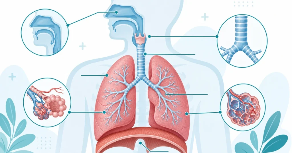
Upper Airway Structures
Air enters the body through the upper airway, which includes several structures that warm, filter, and humidify inhaled air before it reaches the lungs.
Nasal Cavity and Sinuses
The nasal cavity is the first air pathway. It is lined with mucous membranes and tiny hairs (cilia) that trap dust, bacteria, and other particles. The nasal septum divides the cavity into right and left sides. The turbinates (nasal conchae) — superior, middle, and inferior — are bony projections that increase the surface area for warming and humidifying air.
The paranasal sinuses are air-filled cavities in the skull bones connected to the nasal cavity. There are four pairs: frontal (forehead), maxillary (cheekbones), ethmoid (between the eyes), and sphenoid (behind the ethmoid). Sinusitis is one of the most commonly coded respiratory conditions.
Pharynx (Throat)
The pharynx is a muscular tube shared by the respiratory and digestive systems. It has three regions, and knowing which region is involved determines the correct CPT code.
| Region | Location | Key Structures | Common Procedures |
|---|---|---|---|
| Nasopharynx | Behind nasal cavity | Adenoids; eustachian tube openings | Adenoidectomy (42830–42836) |
| Oropharynx | Behind the mouth | Palatine tonsils | Tonsillectomy (42820–42826) |
| Laryngopharynx | Connects to larynx and esophagus | Respiratory/digestive divergence point | Pharyngoscopy codes |
Tonsillectomy and adenoidectomy are commonly coded pharyngeal procedures. Know the difference between tonsillectomy with adenoidectomy (42820) and tonsillectomy alone (42826). Some codes specify age — “under age 12” — so patient age affects code selection.
Larynx (Voice Box)
The larynx sits at the top of the trachea and contains the vocal cords (vocal folds). It has three main functions: protecting the airway during swallowing, producing voice, and serving as an air passage.
Key structures include the epiglottis (a flap that covers the larynx during swallowing to prevent aspiration), the thyroid cartilage (Adam’s apple), and the cricoid cartilage (the only complete ring of cartilage in the airway).
Common conditions include laryngitis, vocal cord paralysis, vocal cord nodules and polyps, laryngeal cancer, croup (in children), and epiglottitis.
Laryngoscopy codes (31505–31579) are organized by approach: indirect (mirror), direct, and flexible fiberoptic. Surgical laryngoscopy codes include biopsy, excision of lesions, injection, and laser ablation. Diagnostic laryngoscopy is bundled into surgical laryngoscopy — the same universal endoscopy bundling rule that applies throughout all body systems.
Trachea (Windpipe)
The trachea is a tube approximately 11 cm long extending from the larynx to the point where it divides into the right and left main bronchi — a landmark called the carina. The trachea is reinforced by C-shaped cartilage rings that keep it open during breathing. The posterior wall is membranous (facing the esophagus), allowing the esophagus to expand during swallowing.
Common conditions include tracheitis, tracheal stenosis (narrowing), tracheomalacia (softening of cartilage), and tracheoesophageal fistula (abnormal connection between trachea and esophagus).
Lower Airway and Lungs
The Bronchial Tree
Below the trachea, the airway branches like an inverted tree, progressively dividing into smaller passages:
Primary (Main) Bronchi: The trachea splits into the right and left main bronchi at the carina. The right main bronchus is shorter, wider, and more vertical than the left. This anatomical fact has a direct clinical consequence: aspirated foreign bodies more commonly lodge in the right bronchus because gravity favors the straighter, more vertical right side.
Secondary (Lobar) Bronchi: Each main bronchus divides into lobar bronchi — three on the right (one for each lobe) and two on the left (one for each lobe).
Tertiary (Segmental) Bronchi: Further division into segmental bronchi, each supplying a bronchopulmonary segment.
Bronchioles: The smallest airways, less than 1 mm in diameter. Terminal bronchioles are the last conducting airways. Respiratory bronchioles begin the gas exchange zone.
Alveoli: Tiny air sacs — approximately 300 million in both lungs — where gas exchange occurs. Oxygen diffuses from the alveoli into the pulmonary capillaries, and carbon dioxide diffuses from the blood into the alveoli for exhalation.
Lung Anatomy
The right lung has three lobes (upper, middle, lower) separated by the oblique and horizontal fissures. The left lung has two lobes (upper and lower) separated by the oblique fissure only. The left lung is slightly smaller because the heart occupies space on the left side of the chest — this anatomical indentation is called the cardiac notch.
| Feature | Right Lung | Left Lung |
|---|---|---|
| Lobes | 3 (upper, middle, lower) | 2 (upper, lower) |
| Fissures | Oblique + horizontal | Oblique only |
| Main bronchus | Shorter, wider, more vertical | Longer, narrower, more horizontal |
| Size | Slightly larger | Slightly smaller (cardiac notch) |
| Lobar bronchi | 3 | 2 |
When coding pneumonia, lung cancer, or other lung conditions, ICD-10-CM requires you to specify which lobe is affected. Codes differentiate between right upper lobe, right middle lobe, right lower lobe, left upper lobe, and left lower lobe. This specificity also applies to surgical procedures. The ICD-10-CM code structure requires this level of anatomical detail throughout the codebook.
Key surgical codes for lung procedures:
| CPT Code | Procedure | What It Involves |
|---|---|---|
| 32440 | Pneumonectomy | Removal of entire lung |
| 32480 | Lobectomy | Removal of one lobe |
| 32484 | Segmentectomy | Removal of a lung segment (less than a lobe) |
| 32505 | Wedge resection | Removal of a small, wedge-shaped piece of tissue |
| 32601–32674 | Thoracoscopy (VATS) | Minimally invasive lung procedures via scope |
Pleura and Surrounding Structures
The lungs are surrounded by protective membranes called the pleura. The visceral pleura is the inner layer directly covering the lung surface. The parietal pleura is the outer layer lining the chest wall, diaphragm, and mediastinum. The pleural space between them normally contains a small amount of serous fluid for lubrication.
| Condition | What It Is | Key Procedure |
|---|---|---|
| Pleural effusion | Fluid in the pleural space | Thoracentesis (32554–32557) |
| Pneumothorax | Air in the pleural space, causing lung collapse | Chest tube (32551) |
| Hemothorax | Blood in the pleural space | Chest tube or thoracotomy |
| Empyema | Pus in the pleural space | Drainage, possible decortication |
| Pleurisy | Inflammation of the pleura | Medical management |
Know the difference between thoracentesis (needle drainage of fluid, 32554–32557) and chest tube insertion (tube thoracostomy, 32551). Thoracentesis codes distinguish between with and without imaging guidance. Pleurodesis (32560) permanently seals the pleural space to prevent recurrent effusions.
The mediastinum is the central compartment of the chest containing the heart, great vessels, esophagus, trachea, and thoracic lymph nodes. Mediastinoscopy (39400) is an endoscopic examination of the mediastinum, often performed for lymph node biopsy in lung cancer staging.
Essential Respiratory Terminology
Respiratory terminology follows the standard medical word-building rules. Mastering these roots, prefixes, and suffixes is essential for interpreting operative reports and clinical notes.
Root Words
The highest-yield respiratory root words are: nas/o and rhin/o (nose), sinus/o (sinus), pharyng/o (pharynx), laryng/o (larynx), trache/o (trachea), bronch/o and bronchi/o (bronchus), pneum/o and pneumon/o (lung or air), pulmon/o (lung), pleur/o (pleura), thorac/o (chest), ox/i (oxygen), and capn/o (carbon dioxide).
Key Prefixes and Suffixes
Prefixes that modify respiratory terms: dys- (difficult — dyspnea), tachy- (fast — tachypnea), brady- (slow — bradypnea), a- and an- (without — apnea, anoxia), hyper- (excessive — hyperventilation, hypercapnia), and hypo- (below normal — hypoventilation, hypoxemia).
Suffixes specific to the respiratory system: -pnea (breathing — dyspnea, apnea, tachypnea, orthopnea), -thorax (chest/pleural cavity — pneumothorax, hemothorax), -ectasis (expansion/dilation — bronchiectasis, atelectasis), and -ptysis (spitting — hemoptysis, or coughing up blood).
Common Respiratory Conditions and Their Coding
Pneumonia
Pneumonia is infection of the lung parenchyma — the alveoli and surrounding tissue. ICD-10-CM codes are organized by causative organism: bacterial (J15.-), viral (J12.-), or organism unspecified (J18.-). The most commonly coded type is J18.9 (pneumonia, unspecified organism).
When the causative organism is identified, code to the specific organism. Aspiration pneumonia (J69.0) has its own code and is sequenced differently — it is caused by inhaling food, liquid, or vomit into the lungs. The ICD-10-CM sequencing rules govern how pneumonia codes interact with manifestation codes and underlying conditions.
COPD (Chronic Obstructive Pulmonary Disease)
COPD includes chronic bronchitis and emphysema. ICD-10-CM distinguishes three key clinical scenarios:
| Code | Scenario | Additional Code Needed? |
|---|---|---|
| J44.0 | COPD with acute lower respiratory infection | Yes — code the infection additionally |
| J44.1 | COPD with acute exacerbation | No — exacerbation is included |
| J44.9 | COPD, unspecified | No |
Asthma
Asthma codes (J45.-) use a two-axis classification: severity (mild intermittent, mild persistent, moderate persistent, severe persistent) and current status (uncomplicated, with acute exacerbation, with status asthmaticus). Status asthmaticus is a life-threatening asthma attack that does not respond to standard treatment — it is a medical emergency and affects reimbursement significantly.
Respiratory Failure
Respiratory failure codes (J96.-) distinguish between acute (J96.0-), chronic (J96.1-), and acute-on-chronic (J96.2-). Each category is further divided by whether it involves hypoxia (low oxygen), hypercapnia (high CO2), or both. Respiratory failure is often a secondary diagnosis that significantly impacts DRG assignment and reimbursement.
When coding respiratory failure with another condition, sequencing depends on which condition prompted the encounter. If the patient presents primarily for respiratory failure, it is the principal diagnosis. If respiratory failure develops during treatment for another condition, the other condition is typically sequenced first. The ICD-10-CM sequencing rules provide the general framework for these decisions.
Key Respiratory Procedure Codes for the CPC Exam
Endoscopy Codes
| CPT Code | Procedure | Key Detail |
|---|---|---|
| 31231 | Diagnostic nasal endoscopy | Visualization of nasal cavity and sinuses |
| 31254 | Partial ethmoidectomy (anterior) | Endoscopic removal of anterior ethmoid cells |
| 31256 | Maxillary antrostomy | Enlargement of maxillary sinus opening |
| 31267 | Frontal sinus exploration | With or without tissue removal |
| 31505 | Indirect laryngoscopy | Using a mirror — simplest approach |
| 31575 | Flexible fiberoptic laryngoscopy | More detailed than indirect |
| 31622 | Diagnostic bronchoscopy | Visualization of tracheobronchial tree |
| 31625 | Bronchoscopy with biopsy | Bronchial wall biopsy — includes diagnostic |
| 31628 | Bronchoscopy with transbronchial biopsy | Biopsy through bronchial wall into lung tissue |
Surgical Codes
Key surgical codes include tracheostomy — planned (31600) and emergency (31603); tube thoracostomy or chest tube insertion (32551); thoracentesis without imaging guidance (32554) and with imaging guidance (32555); and pleurodesis (32560).
Know that 31625 (bronchoscopy with bronchial biopsy) and 31628 (bronchoscopy with transbronchial lung biopsy) are different procedures. A bronchial biopsy samples the bronchial wall itself. A transbronchial biopsy passes through the bronchial wall into the lung parenchyma — it is more invasive and produces a different type of tissue sample. The CPC exam tests this distinction.
CPC Exam Tips for Respiratory System Coding
The eight highest-yield respiratory topics for the CPC exam are:
(1) Airway order: Nasal cavity → pharynx → larynx → trachea → bronchi → bronchioles → alveoli. Questions test whether you can identify where a condition or procedure occurs.
(2) Right lung = 3 lobes, left lung = 2 lobes. This affects pneumonia coding, surgical documentation, and anatomical questions.
(3) Aspiration goes right. Foreign bodies and aspirated material tend to enter the right main bronchus because it is wider and more vertical.
(4) Endoscopy bundling applies. Diagnostic bronchoscopy, laryngoscopy, and nasal endoscopy are all bundled into their surgical counterparts.
(5) Sinus codes are site-specific. Each sinus has its own procedure code — always identify which sinus was operated on.
(6) COPD exacerbation vs. infection. J44.1 (exacerbation) and J44.0 (with infection) are different codes for different clinical scenarios.
(7) Respiratory failure classification. Distinguish acute vs. chronic vs. acute-on-chronic, and hypoxic vs. hypercapnic.
(8) Tracheostomy vs. intubation. Tracheostomy is a surgical procedure creating an external opening. Endotracheal intubation is a tube through the mouth or nose — completely different procedures with different codes.
5 quick questions drawn from this guide. Click an answer to check it — explanations appear as you go.
1. The main airway that branches into the left and right bronchi is the:
2. Gas exchange in the lungs occurs in the:
3. The root “pneum/o” or “pulmon/o” refers to the:
4. The membrane lining the lungs and thoracic cavity is the:
5. “COPD” primarily involves obstruction that is:
Frequently Asked Questions
What is the most commonly tested respiratory anatomy fact on the CPC exam?
That the right lung has three lobes and the left lung has two lobes, and that aspirated foreign bodies tend to lodge in the right main bronchus because it is shorter, wider, and more vertical than the left. Both facts appear in multiple question formats — direct anatomy questions, procedure coding questions involving foreign body removal, and pneumonia coding questions requiring lobe specification.
How do I code COPD with pneumonia?
Use J44.0 (COPD with acute lower respiratory infection) as the primary code, then add a code for the specific type of pneumonia (e.g., J15.9 for unspecified bacterial pneumonia). If the documentation says “COPD exacerbation” without mentioning an infection, use J44.1 instead — no additional infection code. The distinction depends entirely on the clinical documentation.
What is the difference between bronchoscopy with biopsy (31625) and bronchoscopy with transbronchial biopsy (31628)?
CPT 31625 is a biopsy of the bronchial wall — the scope takes a sample from the surface lining of the bronchus. CPT 31628 is a transbronchial lung biopsy — the biopsy instrument passes through the bronchial wall into the lung parenchyma to sample deeper tissue. The transbronchial approach is used when the suspected pathology is in the lung tissue itself, not the bronchial lining. Both include the diagnostic bronchoscopy (31622) — do not report it separately.
How are asthma codes structured in ICD-10-CM?
Asthma codes (J45.-) follow a two-axis pattern: severity level (mild intermittent, mild persistent, moderate persistent, severe persistent) plus current status (uncomplicated, with acute exacerbation, with status asthmaticus). The 4th character identifies the severity, and the 5th character identifies the status. If the severity is not documented, use the “unspecified” codes (J45.90-). Status asthmaticus represents a life-threatening attack and carries significantly higher reimbursement weight.
What does the suffix -ectasis mean and how does it appear in respiratory coding?
The suffix -ectasis means expansion or dilation. In respiratory terminology, it appears in two important conditions: bronchiectasis (chronic abnormal dilation of the bronchi, coded as J47.-) and atelectasis (collapse or incomplete expansion of lung tissue, coded as J98.11). These are opposite conditions — bronchiectasis means the airways are too dilated, while atelectasis means the lung tissue is not expanded enough. Despite sharing the same suffix, they are coded from different ICD-10-CM categories.