Table of Contents
- Overview of the Renal and Urinary System
- Kidney Anatomy in Detail
- The Ureters
- The Bladder
- The Urethra
- Common Renal and Urinary Conditions
- How Renal and Urinary Anatomy Connects to CPT Code Selection
- Kidney Stone Terminology and Coding Context
- The Kidneys' Role in Systemic Disease
- How the CPC Exam Tests Renal and Urinary Anatomy
- Common Mistakes
- Frequently Asked Questions
The renal and urinary system filters waste from the blood, regulates fluid and electrolyte balance, and eliminates liquid waste from the body. For medical coders, this system generates a steady volume of both diagnosis and procedure coding — from chronic kidney disease staging to urinary tract infections to a wide range of urological procedures spanning CPT’s Urinary System subsection. Understanding the anatomy from kidney to urethra, and how each structure’s function connects to specific disease processes, is essential for accurate code selection in this area.
This guide covers the major structures of the renal and urinary system, common conditions affecting each structure, and how this anatomy connects to CPT and ICD-10-CM coding. For related endocrine and metabolic context, see Endocrine System for Medical Coders, and for the broader surgical CPT framework, see Surgery Coding for Beginners.
Overview of the Renal and Urinary System
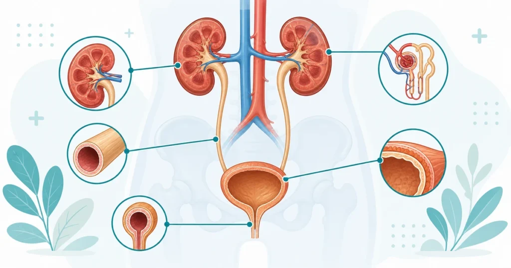
The urinary system consists of four main structures working in sequence: two kidneys, which filter blood and produce urine; two ureters, which carry urine from the kidneys to the bladder; the bladder, which stores urine; and the urethra, which carries urine out of the body during urination.
| Structure | Location | Primary Function |
|---|---|---|
| Kidneys (2) | Retroperitoneal, one on each side of the spine, below the diaphragm | Filter blood, produce urine, regulate fluid/electrolyte balance, produce erythropoietin and renin |
| Ureters (2) | Connect each kidney to the bladder | Transport urine from kidney to bladder via peristalsis |
| Bladder | Pelvic cavity | Stores urine until voiding |
| Urethra | Extends from bladder to external opening | Carries urine out of the body |
Kidney Anatomy in Detail
Each kidney is a bean-shaped organ located retroperitoneally (behind the abdominal cavity lining) on either side of the vertebral column, roughly at the level of the T12 to L3 vertebrae. The right kidney sits slightly lower than the left because of the space occupied by the liver above it.
Internal Kidney Structure
The kidney is organized into three main regions. The renal cortex is the outer region, containing most of the nephrons — the functional filtering units of the kidney. The renal medulla is the inner region, organized into cone-shaped structures called renal pyramids, which contain the tubules that concentrate urine. The renal pelvis is the funnel-shaped structure where urine collects before draining into the ureter.
The Nephron — The Functional Unit
Each kidney contains roughly one million nephrons, the microscopic structures responsible for actually filtering blood and forming urine. Each nephron consists of a glomerulus (a cluster of capillaries where blood filtration begins) surrounded by Bowman’s capsule, followed by a series of tubules (proximal convoluted tubule, loop of Henle, distal convoluted tubule) that reabsorb needed substances and secrete waste, ultimately forming urine that drains into collecting ducts. Glomerular function is directly relevant to coding because glomerular filtration rate (GFR) is the primary measure used to stage chronic kidney disease.
Kidney Functions Beyond Filtration
Beyond filtering blood, the kidneys perform several other critical functions that connect to conditions coded outside the urinary chapter itself. The kidneys produce renin, a hormone that helps regulate blood pressure through the renin-angiotensin-aldosterone system — directly relevant to understanding hypertensive kidney disease. The kidneys also produce erythropoietin, a hormone that stimulates red blood cell production in bone marrow; chronic kidney disease frequently causes anemia because damaged kidneys produce less erythropoietin. The kidneys additionally activate vitamin D and regulate calcium and phosphate balance, which is why chronic kidney disease is often associated with bone and mineral disorders.
The Ureters
The ureters are narrow, muscular tubes, roughly 25 to 30 centimeters long in an adult, that transport urine from each renal pelvis to the bladder through rhythmic muscular contractions called peristalsis. The ureters have three natural areas of narrowing — the ureteropelvic junction (where the ureter meets the renal pelvis), the point where the ureter crosses the iliac vessels, and the ureterovesical junction (where the ureter enters the bladder) — and these narrow points are the most common locations for a kidney stone to become lodged, causing the severe pain characteristic of ureteral colic.
The Bladder
The bladder is a hollow, muscular, expandable organ located in the pelvis that stores urine until voiding. Its wall contains the detrusor muscle, which contracts during urination to expel urine, and the internal lining is made of specialized tissue called transitional epithelium (urothelium) that can stretch significantly as the bladder fills. The point where the ureters enter the bladder and where the urethra exits forms a triangular area on the bladder floor called the trigone, a region of particular clinical significance because it is a common site for bladder tumors and is highly sensitive to irritation, contributing to symptoms like urgency and frequency when inflamed or infected.
The Urethra
The urethra carries urine from the bladder to outside the body and is anatomically different between males and females — a distinction with direct clinical and coding relevance. The female urethra is short, roughly 4 centimeters, which is a key reason women experience urinary tract infections more frequently than men, since bacteria have a shorter distance to travel to reach the bladder. The male urethra is much longer, roughly 18 to 20 centimeters, and passes through the prostate gland before continuing through the penis, meaning male urethral and lower urinary tract conditions are frequently connected to prostate anatomy and pathology.
Common Renal and Urinary Conditions
| Condition | Structure Affected | ICD-10-CM Category |
|---|---|---|
| Chronic kidney disease | Kidney (nephrons) | N18.- |
| Acute kidney injury | Kidney | N17.- |
| Urinary tract infection | Bladder, urethra (lower tract) or kidney (upper tract) | N39.0, N30.-, N10-N12 |
| Nephrolithiasis (kidney stones) | Kidney, ureter | N20.- |
| Benign prostatic hyperplasia | Prostate (affects urethra/bladder outlet) | N40.- |
| Neurogenic bladder | Bladder (nerve control) | N31.- |
Chronic Kidney Disease Staging
Chronic kidney disease (CKD) is staged based on glomerular filtration rate, and ICD-10-CM codes reflect this staging directly — from N18.1 (Stage 1, GFR 90 or greater, with evidence of kidney damage) through N18.5 (Stage 4) and N18.6 (End stage renal disease, requiring dialysis or transplant). Correctly coding CKD requires knowing the documented stage, not simply assigning an unspecified CKD code when a specific stage is available in the record — a frequent source of under-coded specificity.
CKD is also commonly linked to hypertension through combination coding. When documentation establishes both hypertension and CKD, ICD-10-CM assumes a causal relationship under the “with” convention, and category I12 (Hypertensive chronic kidney disease) is used, with an additional code identifying the specific CKD stage. See Hypertension Coding in ICD-10-CM for the complete combination code framework.
Urinary Tract Infections — Upper vs Lower Tract
A urinary tract infection is classified by the location affected. Lower urinary tract infections involve the bladder (cystitis) or urethra (urethritis) and are coded with categories such as N30 (cystitis). Upper urinary tract infections involve the kidney itself, most significantly pyelonephritis (kidney infection), coded from categories N10–N12, and represent a more serious condition with greater risk of complications, including sepsis. Correctly distinguishing upper from lower tract involvement based on documented symptoms and diagnostic findings is essential for accurate code selection.
How Renal and Urinary Anatomy Connects to CPT Code Selection
The Urinary System subsection of CPT (50010–53899) is organized by anatomical structure, generally in the order kidney, ureter, bladder, and urethra, with procedure types (endoscopy, incision, excision, repair) nested within each. A large share of urinary system procedures are performed endoscopically via cystoscopy (a scope passed through the urethra into the bladder) or ureteroscopy (a scope advanced further into the ureter or kidney), and correctly identifying how far the scope was advanced — bladder only versus ureter versus renal pelvis — is often what distinguishes one CPT code from another for what might otherwise seem like a similar procedure.
Procedures for kidney stone removal are a particularly detailed area of the CPT Urinary System subsection, with distinct codes depending on the approach (extracorporeal shock wave lithotripsy, ureteroscopic stone extraction, percutaneous nephrolithotomy) and the specific location of the stone within the urinary tract.
Kidney Stone Terminology and Coding Context
Nephrolithiasis (kidney stones) is one of the most procedurally significant conditions in urology, and coders benefit from understanding the terminology that distinguishes stone location, since ICD-10-CM and CPT both reflect this level of detail. A stone within the kidney itself is termed a renal calculus. A stone that has moved into the ureter is termed a ureteral calculus, and its exact position (proximal, mid, or distal ureter) affects both symptom presentation and the surgical approach used to remove it. Treatment approach depends heavily on stone size and location: extracorporeal shock wave lithotripsy (ESWL) uses focused shock waves from outside the body to fragment smaller stones; ureteroscopy passes a scope up through the urethra, bladder, and ureter to directly visualize and remove or fragment a stone; and percutaneous nephrolithotomy involves a small incision through the back directly into the kidney for larger or more complex stones. Each approach maps to a distinct CPT code, so identifying the documented technique is as important as identifying the stone’s location.
The Kidneys’ Role in Systemic Disease
Because the kidneys interact so closely with the cardiovascular and endocrine systems, renal conditions frequently appear as secondary diagnoses or complications of other systemic diseases rather than as isolated primary conditions. Diabetic nephropathy, discussed in detail in the endocrine system guide, is one of the leading causes of chronic kidney disease and is coded using a diabetes combination code rather than a standalone CKD code. Recognizing these cross-system relationships — diabetes affecting the kidney, hypertension affecting the kidney, kidney disease affecting bone mineral metabolism — helps coders correctly apply the “with” convention and select the appropriate combination codes rather than treating each condition in isolation.
How the CPC Exam Tests Renal and Urinary Anatomy
Pattern 1 — CKD Stage Specificity
The scenario documents a specific CKD stage, and the answer choices include both stage-specific codes and an unspecified CKD code. The correct answer uses the specific documented stage rather than defaulting to unspecified.
Pattern 2 — Upper vs Lower UTI Classification
The scenario describes symptoms and diagnostic findings consistent with either a bladder/urethral infection or a kidney infection, and you must select the code corresponding to the correct anatomical location based on the clinical details provided.
Pattern 3 — Hypertensive CKD Combination Coding
The scenario documents a patient with both hypertension and CKD. The correct answer applies the assumed causal relationship under the “with” convention, using the hypertensive CKD combination code plus an additional code for the specific CKD stage.
Pattern 4 — Endoscopic Procedure Extent
The scenario describes a urinary endoscopic procedure and specifies how far the scope was advanced (bladder, ureter, or renal pelvis). The correct CPT code depends on correctly identifying this extent from the operative documentation.
Common Mistakes
Defaulting to unspecified CKD stage. When a specific GFR-based stage is documented, it must be coded — using an unspecified CKD code when a stage is available under-codes the specificity of the record.
Missing the hypertension/CKD combination code. When both conditions are documented, ICD-10-CM assumes causation under the “with” convention. Coding hypertension and CKD as two unrelated conditions, rather than using the I12 combination code plus stage, is a frequently tested sequencing error.
Confusing upper and lower urinary tract infections. Cystitis (bladder) and pyelonephritis (kidney) are anatomically and clinically distinct conditions with different codes and different clinical severity. Selecting a general “UTI” code when the documentation specifies the kidney is involved under-codes the encounter.
Overlooking male urethral anatomy’s connection to the prostate. Because the male urethra passes through the prostate, many male lower urinary tract symptoms and procedures are tied to prostate pathology (such as benign prostatic hyperplasia), which is a separate but closely related coding consideration.
Selecting a urinary endoscopic code based on entry point alone. The correct code depends on how far the scope was advanced, not merely that a cystoscope was used. A cystoscopy that included ureteral catheterization is coded differently from a bladder-only cystoscopy.
5 quick questions drawn from this guide. Click an answer to check it — explanations appear as you go.
1. The functional filtering unit of the kidney is the:
2. Urine travels from the kidney to the bladder through the:
3. The medical root “nephr/o” and “ren/o” both refer to the:
4. A stone lodged in the ureter is termed:
5. The kidneys contribute to blood pressure regulation via:
Frequently Asked Questions
What are the four main structures of the urinary system?
The urinary system consists of the two kidneys, which filter blood and produce urine; the two ureters, which carry urine from the kidneys to the bladder; the bladder, which stores urine; and the urethra, which carries urine out of the body. Each structure has distinct associated conditions and CPT procedure codes.
What is a nephron?
A nephron is the microscopic functional unit of the kidney responsible for filtering blood and forming urine. Each kidney contains roughly one million nephrons, each consisting of a glomerulus (filtering unit) surrounded by Bowman’s capsule and a series of tubules. Glomerular filtration rate, based on nephron function, is the primary measure used to stage chronic kidney disease.
How is chronic kidney disease staged in ICD-10-CM?
Chronic kidney disease is staged based on glomerular filtration rate, and ICD-10-CM category N18 reflects this staging directly, from N18.1 (Stage 1) through N18.5 (Stage 4) and N18.6 (End stage renal disease). Coders should always use the specific documented stage rather than an unspecified CKD code when the stage is available in the record.
What is the difference between an upper and lower urinary tract infection?
A lower urinary tract infection involves the bladder (cystitis) or urethra (urethritis). An upper urinary tract infection involves the kidney, most significantly pyelonephritis, which is generally more serious and carries a higher risk of complications such as sepsis. These are coded from different ICD-10-CM categories based on the anatomical location documented.
Why does the female urethra length matter clinically?
The female urethra is much shorter than the male urethra, roughly 4 centimeters compared to 18 to 20 centimeters. This shorter distance for bacteria to travel to reach the bladder is a key anatomical reason women experience urinary tract infections significantly more often than men.