Table of Contents
- Why Cardiovascular Anatomy Matters on the CPC Exam
- The Heart's Structure: Chambers and Valves
- Layers of the Heart Wall
- The Cardiac Conduction System
- Coronary Circulation: The Arteries That Feed the Heart Muscle
- Blood Flow Pathway: Systemic and Pulmonary Circulation
- Major Vessels Beyond the Heart
- Building Cardiovascular Vocabulary from Word Parts
- Putting It Together: A Coding Scenario
- Frequently Asked Questions
The cardiovascular surgery section of the CPT manual runs from 33016 to 37799 — one of the longest, densest ranges in the entire Surgery chapter. About ten of the 150 questions on the CPC exam pull from the 30000 series, and most of those are cardiovascular. None of that code range will make sense, though, until you can picture what’s actually happening inside the chest: which chamber pushes blood where, which valve sits between which structures, and which artery is blocked when a chart says “LAD occlusion.” This guide builds that picture, then connects it directly to the terminology and diagnosis codes you’ll see on real charts.
Why Cardiovascular Anatomy Matters on the CPC Exam
Cardiovascular questions show up in two places on the exam: the medical terminology/anatomy section, and the CPT coding section. They overlap constantly. A question might describe an operative note — “aortic valve replaced via median sternotomy” — and ask you to identify the correct approach code, but you can’t even parse the sentence without knowing where the aortic valve sits and what a sternotomy is. Coders who skip the anatomy and jump straight to memorizing code ranges tend to stall out here, because the cardiovascular section uses more Greek- and Latin-derived terms per paragraph than almost any other body system.
📌 CPC Exam Tip: When a practice question gives you an operative note, underline every anatomical term before you look at the code choices. Misreading “epicardial” as “endocardial” — outer surface versus inner lining — is a common trap, and it changes which CPT code applies.
The Heart’s Structure: Chambers and Valves
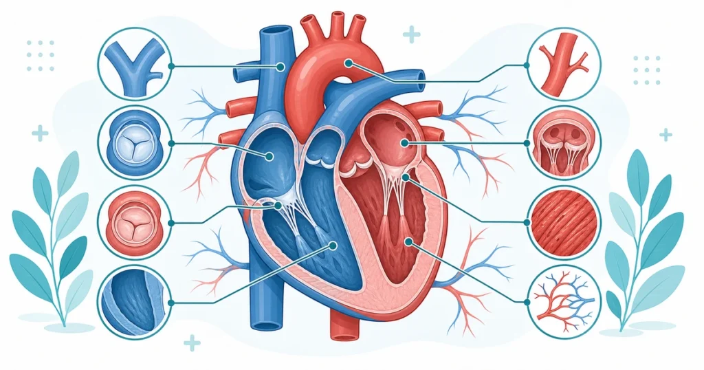
The heart has four chambers, working as two pumps in series.
| Chamber | Location | Function |
|---|---|---|
| Right atrium (RA) | Upper right | Receives deoxygenated blood from the body via the vena cavae |
| Right ventricle (RV) | Lower right | Pumps deoxygenated blood to the lungs via the pulmonary artery |
| Left atrium (LA) | Upper left | Receives oxygenated blood from the lungs via the pulmonary veins |
| Left ventricle (LV) | Lower left | Pumps oxygenated blood to the body via the aorta |
Notice the pattern coders rely on constantly: the right side always handles deoxygenated blood, the left side always handles oxygenated blood. That single fact resolves a lot of confusing terminology on sight.
Four valves keep blood moving in one direction and prevent backflow:
| Valve | Position | Type | Common pathology |
|---|---|---|---|
| Tricuspid | Between RA and RV | Atrioventricular | Tricuspid regurgitation |
| Pulmonary | Between RV and pulmonary artery | Semilunar | Pulmonary stenosis |
| Mitral (bicuspid) | Between LA and LV | Atrioventricular | Mitral valve prolapse, insufficiency |
| Aortic | Between LV and aorta | Semilunar | Aortic stenosis |
Clinical example: An operative note reads, “Patient with severe calcific aortic stenosis underwent aortic valve replacement with a bioprosthetic valve via median sternotomy.” The diagnosis term “stenosis” tells you the valve has narrowed, restricting flow — the ICD-10-CM code is I35.0 (nonrheumatic aortic valve stenosis). The procedure itself, valve replacement, falls in the cardiovascular CPT range and is covered in detail in our guide to heart valve procedure coding.
Layers of the Heart Wall
The heart wall has three layers, and mixing them up is one of the most common mistakes on practice exams:
| Layer | Position | Notes for coders |
|---|---|---|
| Epicardium | Outermost layer | Continuous with the visceral pericardium covering the heart’s surface |
| Myocardium | Middle, muscular layer | Does the actual contracting; site of damage in a myocardial infarction |
| Endocardium | Innermost lining | Lines chambers and valves; site of infection in endocarditis |
The pericardium itself is a separate structure — a double-walled sac surrounding the heart, not a layer of the heart wall. It has a fibrous outer layer and a serous inner layer (parietal and visceral), with the pericardial cavity between them holding a small amount of lubricating fluid. When that fluid builds up abnormally (pericardial effusion) or the sac becomes inflamed (pericarditis), the terminology distinction between pericardium and myocardium becomes the whole ballgame for code selection, since pericardial procedures (33016–33050 range) are billed differently from myocardial procedures.
The Cardiac Conduction System
Electrical terminology trips up a lot of candidates because the structures are tiny and the names are unfamiliar outside of cardiology.
| Structure | Role |
|---|---|
| Sinoatrial (SA) node | The heart’s natural pacemaker; initiates each heartbeat in the right atrium |
| Atrioventricular (AV) node | Delays the signal briefly, letting the atria finish contracting before the ventricles fire |
| Bundle of His | Carries the signal from the AV node into the ventricles |
| Left and right bundle branches | Split the signal toward each ventricle |
| Purkinje fibers | Spread the signal through the ventricular muscle, triggering contraction |
When this system misfires, the resulting conditions generate some of the most frequently tested ICD-10-CM codes in the cardiovascular chapter: I48.0 (paroxysmal atrial fibrillation), I48.91 (atrial fibrillation, unspecified), and I49.01 (ventricular fibrillation). The prefix “fibrillat-” refers to rapid, uncoordinated muscle twitching rather than an organized contraction — once you know that root, you can recognize the family of arrhythmia terms (atrial flutter, ventricular flutter, fibrillation) without memorizing each one separately.
📌 CPC Exam Tip: Pacemaker and defibrillator placement (33206–33249) is one of the most heavily tested code families in this section. Read the operative note for chamber count (single vs. dual) and lead placement (transvenous vs. epicardial) before selecting a code — both details change the answer.
Coronary Circulation: The Arteries That Feed the Heart Muscle
The heart muscle needs its own blood supply, delivered by the coronary arteries, which branch off the aorta just above the aortic valve.
| Artery | Abbreviation | Territory supplied |
|---|---|---|
| Right coronary artery | RCA | Right atrium, right ventricle, SA/AV nodes in most people |
| Left main coronary artery | LM | Splits into the LAD and circumflex |
| Left anterior descending | LAD | Front of the left ventricle and most of the septum |
| Left circumflex | LCX | Lateral and posterior left ventricle |
Clinical example: A cardiac catheterization report notes “90% stenosis of the LAD with normal RCA and LCX.” Translated: the artery feeding the front wall of the left ventricle is almost completely blocked. Left untreated, this pattern of atherosclerotic narrowing is coded as I25.10 (atherosclerotic heart disease of native coronary artery, without angina pectoris). If the blockage progresses to a full blockage and tissue death, the diagnosis shifts to a myocardial infarction code — I21.3 for an ST-elevation MI (STEMI) of unspecified site, or I21.4 for a non-ST-elevation MI (NSTEMI). The distinction between STEMI and NSTEMI comes from the EKG pattern, not the artery involved, and it’s one of the most exam-relevant facts in this entire topic because it changes both the ICD-10-CM code family and, often, the urgency of the CPT-coded intervention that follows.
Blood Flow Pathway: Systemic and Pulmonary Circulation
Coders should be able to trace blood through the entire system in order, because operative notes often describe only one segment and expect you to know what comes before and after it.
Pulmonary circulation (right side of the heart to the lungs and back): right atrium → tricuspid valve → right ventricle → pulmonary valve → pulmonary artery → lungs (gas exchange) → pulmonary veins → left atrium
Systemic circulation (left side of the heart to the body and back): left atrium → mitral valve → left ventricle → aortic valve → aorta → body tissues (gas exchange) → venae cavae → right atrium
Two terms worth nailing down precisely: the pulmonary artery is the only artery in the body that carries deoxygenated blood, and the pulmonary veins are the only veins that carry oxygenated blood. Exam writers like to test this exception because it breaks the “arteries carry oxygenated blood” rule students memorize early and never revisit.
Major Vessels Beyond the Heart
The aorta, the body’s largest artery, divides into named segments as it travels from the heart: the ascending aorta, aortic arch, descending thoracic aorta, and abdominal aorta. Each segment matters for vascular CPT coding, since procedures on arteries and veins outside the heart itself fall in the 34001–37799 range — a separate sub-range from the heart and pericardium procedures (33016–33999). Major branches off the aortic arch include the brachiocephalic trunk, left common carotid artery, and left subclavian artery, each supplying a different region (head, neck, and upper limb, respectively).
On the venous side, the superior vena cava drains blood from the head, neck, and upper body, while the inferior vena cava drains the lower body — both emptying into the right atrium. Confusing “superior” and “inferior” here is a frequent terminology slip; for a refresher on directional terms generally, see our guide to anatomical planes and body positions.
Building Cardiovascular Vocabulary from Word Parts
Most cardiovascular terms break down into a handful of recurring roots, prefixes, and suffixes. Recognizing these lets you decode unfamiliar terms on the exam instead of guessing.
| Word part | Meaning | Example term | Translation |
|---|---|---|---|
| cardi/o | heart | cardiomegaly | Enlarged heart |
| angi/o | vessel | angiography | Imaging of vessels |
| ather/o | fatty plaque | atherosclerosis | Hardening from plaque buildup |
| brady- | slow | bradycardia | Slow heart rate |
| tachy- | fast | tachycardia | Fast heart rate |
| -itis | inflammation | pericarditis | Inflammation of the pericardium |
| -megaly | enlargement | cardiomegaly | Enlarged heart |
| -sclerosis | hardening | arteriosclerosis | Hardening of arteries |
| -stenosis | narrowing | aortic stenosis | Narrowing of the aortic valve |
For a broader walkthrough of how prefixes and suffixes combine across every body system, see medical terminology word roots, prefixes, and suffixes.
📌 CPC Exam Tip: When a term combines two roots you recognize — like “cardiomyopathy” (cardi/o + myo + -pathy, disease of the heart muscle) — break it into pieces before guessing the definition from context. Exam distractors are often built by swapping one root for a similar-sounding one.
Putting It Together: A Coding Scenario
A 64-year-old presents to the ED with crushing chest pain radiating to the left arm. EKG shows ST elevation in the anterior leads. The patient is taken emergently to the cath lab, where catheterization reveals 95% occlusion of the LAD. A drug-eluting stent is placed.
Walking through the terminology: “ST elevation” plus “anterior leads” points to a STEMI affecting the front wall of the left ventricle — territory supplied by the LAD, which matches the catheterization finding. The diagnosis code is I21.02 (ST elevation myocardial infarction involving the left anterior descending coronary artery) once the specific vessel is documented, rather than the unspecified-site code I21.3. The procedure — percutaneous coronary intervention with stent placement — is reported separately using codes from the cardiac catheterization and intervention range, which is covered in depth elsewhere on the site. Notice how every step depended on connecting an anatomical term (LAD, anterior wall) to a diagnosis code (I21.02) before the procedure code could even be selected. That sequence — anatomy first, diagnosis second, procedure third — is the backbone of accurate cardiovascular coding.
Hypertension follows the same pattern. A chart noting “essential hypertension” alone codes to I10, but once you see documentation of hypertensive heart disease, the code shifts to the I11 family. Our guide on hypertension coding in ICD-10 covers those distinctions, and pathophysiology for medical coders walks through how disease processes like atherosclerosis connect anatomy to the diagnosis codes that follow from it.
5 quick questions drawn from this guide. Click an answer to check it — explanations appear as you go.
1. The human heart has how many chambers?
2. The valve between the left atrium and left ventricle is the:
3. The three layers of the heart wall, inner to outer, are:
4. The coronary arteries supply blood to:
5. The largest artery in the body is the:
Frequently Asked Questions
What are the four chambers of the heart, and what does each one do?
The right atrium and right ventricle receive deoxygenated blood from the body and pump it to the lungs. The left atrium and left ventricle receive oxygenated blood from the lungs and pump it to the rest of the body. The right side always handles deoxygenated blood; the left side always handles oxygenated blood.
What is the difference between the epicardium, myocardium, and endocardium?
These are the three layers of the heart wall, from outside to inside. The epicardium is the outer layer, continuous with the pericardial sac. The myocardium is the thick, muscular middle layer that contracts to pump blood. The endocardium is the thin inner lining that contacts the blood directly inside the chambers and valves.
Which coronary artery supplies the front wall of the left ventricle?
The left anterior descending artery (LAD), a branch of the left main coronary artery, supplies the anterior wall of the left ventricle and most of the interventricular septum. Blockages here are a frequent cause of anterior-wall myocardial infarctions.
What is the difference between a STEMI and an NSTEMI?
Both are types of myocardial infarction, distinguished by EKG findings rather than which artery is involved. A STEMI shows ST-segment elevation, generally indicating a complete coronary artery blockage. An NSTEMI lacks ST elevation and typically reflects a partial blockage. ICD-10-CM codes these separately — the I21 category for STEMI by site, and I21.4 for NSTEMI.
What does the SA node do, and why is it called the heart’s natural pacemaker?
The sinoatrial (SA) node is a small cluster of specialized cells in the right atrium that generates the electrical signal initiating each heartbeat, without needing input from the nervous system. That signal travels through the AV node, the bundle of His, and the Purkinje fibers to coordinate atrial and ventricular contraction.